

Retroflexed Uterus
Physicians use the term “retroflexed uterus” to describe a condition in which the uterus is tilted backward within the pelvis. At first glance, this might seem dramatic, but it affects approximately 10% of women [1]. However, what are the implications of having a tilted or kinked uterus, and how does this position relate to endometriosis?
Today, we will delve into the retroflexed uterus, exploring its effects and treatment options.
What a “Normal” Uterus Looks Like
The uterus, a vital organ in your body, typically assumes a pear-shaped form. Nestled within the pelvic region, it resides snugly between the rectum and the bladder. This positioning is maintained with ligaments, known as retaining ligaments, constructed from connective tissue.
These retaining ligaments, though essential, allow for a degree of flexibility in the uterus’s position. In other words, the uterus is not rigid; it can adapt to internal and external factors. This adaptability is crucial, especially during pregnancy.
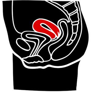
Under usual circumstances, the uterus is slightly bent forward, angling toward your belly button. This positioning, referred to as anteversion, is entirely normal. If you were to examine an image of the uterus, you would observe this natural forward curvature [2].
Forward Tilted Uterus:
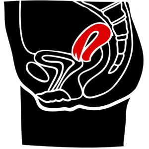
Backward Bending Uterus:
What Is a Retroflexed Uterus?
Defining the Norm: Within the realm of human anatomy, variations are not uncommon, extending to the positioning of the uterus. While conventionally, the uterus tilts forward, it is essential to recognize that it may incline in the opposite direction in some individuals.
When the uterus is tilted or bent towards the spine, it is referred to as a retroflexed uterus. In certain instances, it may even rest against the rectum.
Retroflexed uteri can manifest in two distinct forms: the mobile retroflexio uteri mobilis and the immobile retroflexio uteri fixata. As the names imply, one form allows for mobility of the uterus, while the other locks it in a fixed position [1].
Good to Know!
When comparing the flexible and inflexible forms, it is observed that symptoms are more prevalent in cases of inflexible retroflexio uteri fixata.
Inverted Uterus: Symptoms
A critical point is that having an upside-down uterus does not automatically imply illness. Many women with a retroflexed uterus do not encounter any symptoms.
Nevertheless, not all patients fall into this category, as the altered position within the pelvis can impact neighboring organs. For instance, if the uterus rests on the bowel, it may disrupt the digestive process.
The following symptoms can be associated with an inverted uterus:
- Low back pain
- Constipation
- Painful menstrual bleeding (dysmenorrhea)
- Painful sexual intercourse (dyspareunia)
Retroflexed Uterus: Implications for Childbearing and Pregnancy
In essence, having a retroflexed uterus should not inherently impede one’s ability to conceive. Sperm can naturally reach their intended destination for fertilization. However, if an extended period passes without a successful pregnancy, it becomes crucial to investigate the underlying factors associated with this uterine position. Conditions such as endometriosis, for instance, could potentially hinder conception.
If you are already pregnant, there is a high likelihood that the uterus will spontaneously reposition itself. It very rarely remains inverted. As the uterus expands during pregnancy, it can exert pressure on the symphysis, which, on rare occasions, might lead to incomplete bladder emptying [1].
Uterine Sacculation: A Rare but Serious Complication
Uterine sacculation is a rare yet significant complication that can arise when a retroflexed uterus fails to reposition itself during pregnancy. This condition causes abnormal stretching of the lower uterine segment, encompassing the urinary bladder, cervix, and uterus, potentially leading to a bulge [3].
The following prominent symptoms serve as indicators of uterine sacculation: [3]
- Abdominal pain
- Constipation
- Urinary dysfunction
Timely diagnosis by medical professionals is crucial before childbirth to mitigate pronounced disease symptoms and potential complications. Magnetic resonance imaging can provide valuable insights into the anatomical location, aiding the planning of a cesarean section [3].
It is worth noting that uterine sacculation often necessitates a cesarean section to prevent uterine tears (uterine rupture) and injuries to the bladder or cervix during the surgical procedure [3].
Good to Know!
The likelihood of developing sacculation, characterized by a bulge in the uterus, is estimated to be approximately 1 in 3,000 cases [4].
Causes of Retroflexed Uterus
It is essential to differentiate between an overturned uterus’s flexible and inflexible forms. In the case of a relaxed retroflexed uterus, it can occur as early as childhood. Physicians may also observe this phenomenon following pregnancy when the uterus ultimately fails to return to its original position. Another potential cause is the atrophic uterus, which can develop during postmenopause [1].
It is worth noting that uterine atrophy, a normal aging process, results from the changing hormonal profile in a woman’s body, rendering the uterus unnecessary for reproduction.
In contrast, an immobile retroflexed uterus is often associated with inflammatory processes, prior surgeries, or endometriosis. These factors can lead to the formation of adhesions between the uterus and the rectum [1].
Endometriosis and Retroflexed Uterus: The Connections
Endometriosis, a condition in which endometrial tissue grows outside the uterine cavity, is characterized by a range of symptoms, with abdominal pain as a primary concern. These symptoms frequently manifest during menstruation or in association with sexual intercourse, sometimes accompanied by vomiting, diarrhea, or nausea [5].
The presence of unwanted endometrial tissue can lead to adhesions, resulting in the fixation and retroflection of the uterus.
Physicians often utilize the retroflexed uterus as a potential indicator of endometriosis. Direct visualization of adhesions in the pelvis can be challenging, so gynecologists rely on “soft markers,” which are indirect signs suggesting the presence of endometriosis-related adhesions. When fixed, tilted backward, and bent, the retroflexed uterus resembles a question mark. This unique shape can indicate the presence of endometriosis lesions in the pelvis [6].
Additionally, during ultrasound examinations, if the uterus exhibits no movement relative to surrounding tissues, it may signal a “frozen pelvis,” a condition often associated with severe endometriosis.
Diagnosing a Retroflexed Uterus
Given that many women with a retroflexed uterus remain asymptomatic, its diagnosis is often an incidental finding during routine gynecological check-ups. Your gynecologist may inform you unexpectedly that your uterus is retroflexed.
Gynecologists may employ transvaginal 2-D ultrasound or palpation (vaginal or rectal) to detect and confirm the presence of a retroflexed uterus.
If endometriosis is suspected, physicians will specifically examine the position of the uterus. In cases of deep infiltrating endometriosis, the uterus frequently presents in a retroflexed state [7].
For adenomyosis, a condition where endometrial glandular tissue infiltrates the uterine muscle tissue, the question mark appearance on ultrasound is also a diagnostic clue [7].
Treating a Retroflexed Uterus
If you experience no symptoms, a retroflexed uterus typically does not require treatment. However, if you experience pain or encounter difficulty with urination, several treatment options are available.
In cases of symptomatic retroflexed uteri, physicians may attempt manual repositioning, which involves using their hands to move the uterus into the correct position. Alternatively, a specialized device called the Smith-Hodge pessary can be inserted to realign the uterus [1].
If manual therapy proves effective in alleviating symptoms, you can consult your gynecologist to determine whether surgery is viable. Surgical intervention may involve shortening a retaining ligament (such as Ligg. rotunda or Ligg. sacrouterina) [1, 8].
When endometriosis adhesions are responsible for the retroflexed uterus, surgery can also be considered to mobilize the uterus, which entails breaking adhesions and removing the endometriosis. It is important to note that endometriosis lesions may recur following surgery [9].
In a Nutshell
The uterus is oriented toward the abdominal wall in the typical anatomical position. However, a retroflexed uterus is characterized by its backward tilt, affecting approximately 10% of women. Importantly, it is not inherently a pathological condition, as many women with this anatomical variation live without associated symptoms.
Nonetheless, in some instances, a retroflexed uterus can lead to discomfort, including low back pain, constipation, painful intercourse, or dysmenorrhea (painful menstruation). Treatment becomes necessary only when such symptoms are present. Doctors can reposition the uterus manually or employ a pessary to alleviate this discomfort. An alternative approach involves shortening the retaining ligaments.
It is worth noting that a retroflexed uterus can result from various factors. It may be present from birth, occur due to the natural regression of the uterus with age, or develop in response to inflammation or adhesions. Additionally, retroflexed uteri can be associated with conditions such as endometriosis.
If you are curious about other symptoms and concerns related to endometriosis, consider downloading the Endo-App to access expert knowledge and insights.
References
- Diedrich, Klaus. Gynecology and obstetrics (Springer textbook) (German Edition) (p.165). Springer Berlin Heidelberg. Kindle version.
- Pschyrembel Online | Uterus
- Linder N, Tauscher A, Borte G. Sacculation of the uterus–A rare but ominous complication in pregnancy [Sacculation of the uterus – a rare but ominous complication in pregnancy]. Rofo. 2015 Jan;187(1):57-8. English. doi: 10.1055/s-0034-1366598. epub 2014 Aug 29. PMID: 25171503.
- Thieme E-Journals – Obstetrics and Gynecology The fixed retroflexed uterus in pregnancy with development of posterior sacculation – a case report.
- Health information: Endometriosis
- Di Donato N, Bertoldo V, Montanari G, Zannoni L, Caprara G, Seracchioli R. Question mark form of uterus: a simple sonographic sign associated with the presence of adenomyosis. Ultrasound Obstet Gynecol. 2015 Jul;46(1):126-7. doi: 10.1002/uog.14750. epub 2015 May 27. PMID: 25486912.
- German Society of Gynecology and Obstetrics: guideline program. Diagnosis and therapy of endometriosis. August 2020.
- Pschyrembel Online | Retroflexio uteri.
- Diedrich, Klaus. Gynecology and obstetrics (Springer textbook) (German Edition) (p.166). Springer Berlin Heidelberg. Kindle version.

- Pharmaceutical Companies and Their Influence on Scientific Research - 7. October 2023
- Identification of a Genetic Factor Linked to Endometriosis Development and Potential Therapeutic Targets - 6. October 2023
- Identification of a Genetic Factor Linked to Endometriosis Development and Potential Therapeutic Targets - 6. October 2023