Procedure of Laparoscopy – Explained Step by Step
Laparoscopy is a gentle procedure used for the diagnosis and treatment of endometriosis. If symptoms, palpation, ultrasound and, if necessary, magnetic resonance imaging lead to the suspected diagnosis of “endometriosis”, further diagnostics should be performed by laparoscopy according to the guideline program [1]. The procedure is performed on an outpatient or inpatient basis, depending on the extent of the surgery, so in some cases you can even go home a few hours after the procedure. If endometriosis lesions are removed, the length of your stay depends on the extent of the surgery. In the following, you will learn how the laparoscopy works.
Endometriosis: The most important Facts about the Disease
Endometriosis is one of the most common diseases affecting women of reproductive age. It is estimated that around 1.7 million women (in Germany alone) suffer from endometriosis. Endometriosis is characterized by the fact that cells similar to those from the lining of the uterus (endometrium) grow outside the uterus in so-called endometriosis lesions. These can be found in the pelvis, but also in the intestines, bladder, kidneys, peritoneum and even the lungs. The most common symptoms of endometriosis include bleeding disorders, abdominal pain, dizziness and reduced fertility.
Abdominal Endoscopy: Basic Information and Advantages of the Procedure
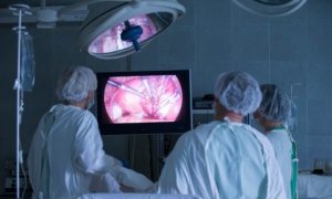
Abdominal endoscopy, called laparoscopy by specialists, is also known as “keyhole surgery”. This procedure can be used both in the field of diagnostics, i.e. to clarify a suspected diagnosis, and for surgical interventions. Unlike traditional abdominal surgery, the abdominal wall needs to be cut only minimally, and large abdominal incisions are not required. In most cases, the incisions are about 1 cm long. This reduces the risk during and after surgery and patients recover more quickly. Finally, there are no long, noticeable scars, which is a cosmetic advantage.
The laparoscopy is sometimes performed on an outpatient basis, which means that patients come to the clinic or care center the morning of the procedure and are discharged home a few hours after the procedure.
This depends on the extent of the internal wounds, i.e. the extent of the surgery. If only a look is made and, for example, no endometriosis or only small superficial foci are found, the surgery can be performed on an outpatient basis.
If endometriosis is found, a hospital stay is often involved. The length is very individual and also relevant from the location of the endometriosis foci as well as the organs involved in the surgery.
Laparoscopy is performed under general anesthesia. Laparoscopy can be used for diagnostic purposes as well as for sampling and surgery. The extent of the operation will be discussed with you by a doctor during the consultation. Here, all eventualities are gone through, since the final operation naturally depends on the exact picture of the endometriosis that is presented to the surgeon in the laparoscopy.
In this article you will learn:
- The advantages of laparoscopy,
- Preparation and educational discussion,
- what preparations are made in the operating room,
- the procedure of laparoscopy in 7 steps,
- behavioral instructions and aftercare following the operation.
Before the Procedure: Preparation for Laparoscopy
A few days before the operation, a preliminary discussion takes place in the clinic or care center where the operation is to be performed. On the one hand, this includes an informative talk about the operation itself and, on the other hand, a preliminary talk with the anesthetist. You will also receive an information sheet containing all the important information about the operation. This may differ in some points from the following presentation. This step-by-step illustration is only a general representation of how an abdominal endoscopy is performed to diagnose endometriosis.
The following points in particular are clarified in the course of the clarification discussion:
- Medications: This is where we discuss which medications need to be discontinued and at what interval prior to surgery, and which can/should continue to be taken. In particular, blood-thinning medications, including over-the-counter medications such as aspirin, must be discontinued as a rule. Otherwise, heavy bleeding can occur during the procedure, which can lead to complications.
- Food intake: No food should be consumed for at least six hours prior to surgery (the exact guidelines are provided by the respective hospital/care center). This includes soups and sweets. Drinks such as fruit juices, milk or alcohol may also no longer be consumed. Smoking and chewing gum should also be avoided. This is usually discussed by the anesthesiologist, since muscle-relaxing drugs are used for anesthesia and surgery. In this condition, the stomach should be empty to avoid vomiting and related complications.
- During the educational interview, the removal of endometriosis lesions is usually also discussed in various points. If endometriosis is found on the bowel or bladder and is removed, there are additional aspects and surgical procedures as well as risks that need to be discussed, no matter how likely this is in your case. For example, even if endometriosis is not suspected in your bowel or bladder, the surgeon must know how to proceed if something is found there. Therefore, you will also go through these scenarios in the explanations. In concrete terms, this means that the risks are explained to you and you then discuss together what should be done and what should first be discussed again after the operation.
Step-by-Step: Laparoscopy Unveiled
[3]
Operation Preparation:
Before the actual laparoscopic procedure, the induction of anesthesia marks the initial phase. Patients are gently relocated in their beds to an anteroom adjoining the operating room, where they are then transferred to a specialized couch. This mobile platform will transport them to the area where anesthesia will be administered. This designated space may vary in size, sometimes within the operating room itself, but more commonly in a separate anteroom to shield patients from witnessing the surgical environment while awake. During this preparation, monitoring equipment, including devices for tracking pulse and blood pressure, is thoughtfully affixed, ensuring close observation of vital signs. Furthermore, venous access is skillfully established to facilitate the delivery of intravenous fluids and medications. The choice of anesthesia procedure rests with the capable anesthesiologist, who meticulously selects the most appropriate method and skillfully administers the medication.
In accordance with the specific clinic’s practices and the estimated duration of the laparoscopy, a bladder catheter may also be inserted before the commencement of the surgery. The purpose of this catheter is to facilitate the drainage of urine during the operation. Although the placement of a catheter may cause slight discomfort, it is generally deemed painless. In some instances, the catheter insertion is deferred until the anesthesia has taken full effect
Step 1: Insertion of a Special Needle (Hollow Needle)
As the laparoscopic procedure commences, the first pivotal step involves creating access to the abdominal cavity through the abdominal wall. Following meticulous disinfection of the skin, a specialized hollow needle is employed to pierce through the layers of the abdominal wall, encompassing the skin, fatty tissue, and muscles. This crucial entry point is typically established in the lower vicinity of the navel, adhering to standard practice.
Step 2: Introducing Gas into the Abdominal Cavity
With the access established, the next critical phase involves the introduction of gas into the abdominal cavity. Typically, between two and four liters of CO2 gas are delicately infused, accounting for variations in the patient’s height, weight, and anatomical condition. The gas ensures the elevation of the abdominal wall, facilitating the smooth insertion of devices and instruments. It also enhances the visualization of internal organs. To maintain safety and ideal conditions, continuous monitoring of the abdominal cavity’s pressure is in place. This vigilant oversight ensures that the gas remains within the correct amount, preventing any potential risks associated with excessive pressure.
Step 3: Insertion of Guide Sleeve and Laparoscope
The puncture needle is seamlessly substituted by a guide sleeve (trocar). Crafted from either plastic or metal, this sleeve boasts a diameter ranging from 5 to 12 mm. A built-in valve effectively ensures that the gas remains contained within the abdominal cavity, preventing any unintended leakage. Beyond gas containment, the guide sleeve serves another crucial function – it stabilizes the opening in the abdominal wall. This stability enables the smooth insertion of a laparoscope into the abdominal cavity. The laparoscope, an indispensable instrument in laparoscopy, typically encompasses a camera, a light source, and a lens magnification system positioned at the end of a slender tube.
In essence, the laparoscope resembles a contemporary camera, equipped with a magnifying glass and illuminating source.
Subsequently, to comprehensively examine all areas of the abdomen, an additional instrument is introduced through a fresh incision. This instrument allows for gentle organ displacement, providing an unobstructed view and enabling meticulous scrutiny of every aspect of the abdominal region.
Step 4: Positioning the Patient
Throughout the operation, a subtle tilt of the operating table places the patient in a carefully inclined position with the head directed downward. This intentional adjustment not only ensures the patient’s secure fixation to the operating table but also effectively prevents any slippage, prioritizing safety during the surgical procedure. This overhead positioning causes the gradual upward movement, shifting towards the head, rendering the organs within the small pelvis accessible to the surgical team.
Step 5: Commencement of the Actual Examination
With the patient appropriately positioned, the surgeon proceeds to embark on the actual examination, involving a comprehensive exploration of the individual organs within the abdominal cavity. This involves a 360-degree rotation of the camera, enabling the visualization of the entire abdominal cavity, up to the diaphragm.
Step 6: Extension of the Procedure for Further Diagnostics
If tissue samples are also to be taken or other minor procedures are to be performed, the laparoscopic procedure may require the creation of additional access points. These additional access points are carefully established through the abdominal wall, typically in the pubic area or in proximity to existing scars on the abdomen. Once the abdominal wall is opened at these designated points, trocars are introduced to provide entry. Through these trocars, various specialized instruments, such as grasping, cutting, or other tools, can be inserted. This expanded access allows for the retrieval of tissue samples, enabling the examination of potential endometriosis lesions or other pathologies.
In more complex scenarios, major surgical procedures, such as partial bladder resection or the removal of large endometriosis lesions, can also be performed using laparoscopy. It is essential not to underestimate the significance of these interventions, due to the smaller external scars. The expertise of the surgeon ensures that the appropriate measures are taken to address the specific surgical requirements.
Step 7: Completion of the Procedure
With all examinations and necessary interventions accomplished, meticulous attention is directed toward the abdominal cavity to ensure the absence of any potential bleeding. Should any bleeding be detected, the skilled surgical team promptly addresses it, often employing laparoscopic techniques to control and halt the bleeding effectively. Additionally, to aid in wound healing and drainage of secretions or blood, a drain may be carefully positioned.
Usually, however, following a thorough examination, the surgical devices utilized throughout the laparoscopy are gently removed and the openings in the abdominal wall are meticulously sutured and dressed with adhesive plasters. Any residual gas present in the abdomen will naturally disperse over time.
Typically, the entire laparoscopic procedure is concluded within a brief duration of 10 to 20 minutes. However, if supplementary examinations or procedures are performed during the laparoscopy, the total time period may extend accordingly.
After the Procedure: Behavioral Instructions and Post-Treatment
Following the procedure, the patient remains in the recovery room, where careful monitoring ensues. It is worth noting that the time spent in the operating area may be extended due to transportation and waiting times. Thus, relatives should be aware that longer absences from the ward might be necessary while they await the patient’s return.
Once the effects of anesthesia have subsided, and the patient’s vital signs, including pulse and blood pressure, stabilize, outpatients are permitted to return home. However, it is crucial that an adult accompanies them and stays with them for the first 24 hours, ensuring immediate assistance in case of any emergencies.
As for inpatients, they recover on the ward under close observation and care.
In the first few days following the procedure, it is common to experience pain in the abdomen and the shoulder region due to the presence of introduced gas. If these discomforts intensify or if a fever develops, it is essential to promptly contact a doctor or seek assistance from an emergency doctor.
For the first three to four weeks after the surgery, it is advised to refrain from lifting loads exceeding five kilos (specific advice from the doctor should be followed). During this recovery period, engaging in sports activities should also be avoided.
Showering is usually permissible immediately after the operation, with the use of waterproof plasters to protect the wounds. After 1–2 days, the wounds are sufficiently closed to withstand water running over them. However, direct rubbing with soap should be avoided during this period. These guidelines generally apply to standard laparoscopies. As always, it is crucial to discuss these recommended actions with the responsible surgeon, ensuring tailored and individualized post-operative care and instructions.
Summary:
Laparoscopy, also known as diagnostic laparoscopy, serves both diagnostic and treatment purposes for endometriosis. This minimally invasive surgical technique involves inserting instruments through small openings in the abdominal wall to view the abdominal organs and obtain samples for verifying the suspected diagnosis of “endometriosis.” The procedure is usually performed on an outpatient basis but under general anesthesia. According to the guideline program, laparoscopy is recommended for the diagnosis of endometriosis, after prior sonography and magnetic resonance imaging, if necessary.
Would you like to learn more about the preparation and procedure of an endometriosis operation? Just download the Endo-App and benefit from the knowledge of our endometriosis experts.
References
- https://www.awmf.org/uploads/tx_szleitlinien/015-045l_S2k_Diagnostik_Therapie_Endometriose_2020-09.pdf
- Andreas D. Ebert; Endometriosis, A Guide for the Practice; De Gruyter.
- https://www.operieren.de/e3224/e10/e886/e898/e900/
- https://www.uksh.de/Frauenklinik_Luebeck_MIC/Laparoskopische+Aufkl%C3%A4rung.html
- https://www.ag-endoskopie.de/bauchspiegelung
- Endometriosis and Migraine: What is the Connection Between the Two Conditions? - 8. October 2023
- Endometriosis and Migraine: What is the Connection Between the Two Conditions? - 8. October 2023
- Can I Inherit Endometriosis? - 7. October 2023


